
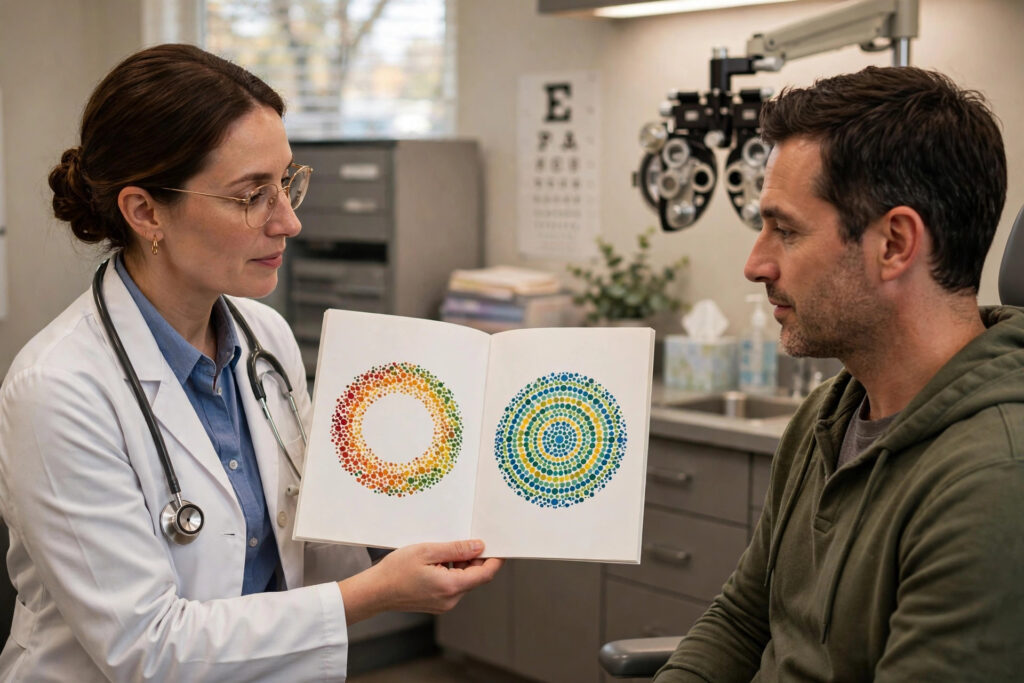
The Ishihara color vision test is one of the most recognizable eye tests in existence: those circular plates filled with colored dots, hiding a number that some people see instantly and others cannot find at all. Developed over a century ago, it remains the standard screening tool for red-green color vision deficiency worldwide.
This guide explains how the Ishihara test works, what the plates are designed to reveal, how results are scored, what color blindness actually means for daily life, and which other tests your doctor might use.
Table of Contents
- What the Ishihara Test Is
- How the Plates Work
- Taking the Test
- How Results Are Scored
- Types of Color Vision Deficiency
- Color Blindness in Daily Life
- Other Color Vision Tests
What the Ishihara Test Is
The Ishihara test was created in 1917 by Dr. Shinobu Ishihara, a professor at the University of Tokyo, originally to screen military recruits. It consists of a book of plates — typically 38 in the full edition — each containing a circle of randomized colored dots. Within the dot pattern, dots of a contrasting color form a number, a winding path, or nothing at all.
People with normal color vision see the hidden figures easily. People with red-green color vision deficiency see different numbers, see nothing, or trace the wrong path — because the colors that distinguish the figure from the background fall within the range they cannot tell apart. The test is fast, inexpensive, and requires no special equipment beyond the plate book and good lighting.
Despite its age, the Ishihara test remains remarkably effective as a screening tool. It is used in schools, occupational health exams, driver’s license assessments in some countries, and as part of routine eye health screenings for children and adults.
How the Plates Work
Each plate exploits the specific color confusions of red-green deficiency. The background dots and figure dots are chosen so that they differ mainly in the red-green dimension while varying randomly in brightness and saturation. This prevents anyone from “cheating” by using brightness cues — you genuinely must distinguish the hues to see the figure.
The plates come in several designs. Demonstration plates are visible to everyone and confirm the patient understands the task. Transformation plates show one number to people with normal vision and a different number to those with color deficiency. Vanishing plates show a figure only to those with normal vision. Hidden digit plates do the reverse — only color-deficient viewers see the number. Diagnostic plates distinguish protan (red-weak) from deutan (green-weak) defects.
Proper illumination is critical. The plates must be viewed under natural daylight or a standardized daylight illuminator — ordinary yellowish indoor lighting shifts color perception and can produce false failures. This is one reason online Ishihara tests on uncalibrated screens are unreliable for diagnosis.
Taking the Test
You will sit in well-lit conditions and view the plates at a comfortable reading distance, usually about 75 centimeters. The examiner shows each plate for a few seconds — typically under five — and asks what number or path you see. There is no penalty for guessing, and you should answer with the first thing you perceive rather than studying the plate.
Both eyes are usually tested together first; if a deficiency is found, each eye may be tested separately to check whether the defect is equal in both eyes. Asymmetric color vision loss can indicate an acquired problem — such as optic nerve disease — rather than the inherited form.
The test is quick and entirely comfortable. Children as young as four or five can take a simplified version using winding-path plates instead of numbers. Parents can learn more about age-appropriate vision testing for children to know what to expect at pediatric exams.
How Results Are Scored
Scoring is straightforward: the examiner counts how many plates you read correctly. Missing a small number of plates can be normal — even people with typical color vision occasionally misread one under imperfect conditions. As a general rule, correctly reading the vast majority of plates indicates normal color vision, while systematic errors across many plates indicate deficiency.
The pattern of errors matters as much as the count. Errors concentrated on transformation and vanishing plates suggest red-green deficiency; the diagnostic plates then indicate whether it is protan-type (red) or deutan-type (green) and roughly how severe. Complete failure on nearly all plates suggests a severe defect.
Important caveat: the Ishihara test screens for red-green deficiency but does not reliably detect blue-yellow defects, which are rarer and usually acquired through eye disease, medication, or aging. A “pass” on Ishihara does not rule out all color vision problems — which is why occupational screenings for pilots and electricians often add supplementary tests.
Types of Color Vision Deficiency
Red-green deficiency is by far the most common type, affecting about 1 in 12 men and 1 in 200 women of Northern European ancestry. It is X-linked: the genes for red and green cone pigments sit on the X chromosome, so men (with one X) are affected far more often than women (who need the variant on both X chromosomes). Deuteranomaly (green-weak) is the most common subtype, followed by protanomaly (red-weak); complete dichromacy (protanopia, deuteranopia) is less common.
Blue-yellow deficiency (tritan defects) is rare when inherited but can be acquired through cataracts, glaucoma, macular degeneration, diabetes, or medications. Because acquired defects often affect blue-yellow perception first, new color vision problems in adulthood deserve a full eye examination rather than being dismissed as “just color blindness.”
Total color blindness (achromatopsia) — seeing only in shades of gray — is extremely rare, affecting roughly 1 in 30,000 people. It is usually accompanied by light sensitivity, poor visual acuity, and involuntary eye movements, and it is a distinct condition from common red-green deficiency.
Color Blindness in Daily Life
For most people with red-green deficiency, daily life is barely affected. The condition is a difference in color perception, not a loss of vision — acuity is typically normal. Common challenges include distinguishing ripe from unripe fruit, matching clothes, reading certain charts and maps, and identifying some traffic or wiring color codes — though traffic lights are also distinguished by position and brightness.
Certain careers have color vision requirements: commercial pilots, air traffic controllers, electricians, and some military roles. If your child is diagnosed, it is worth knowing early so career planning can account for it. Schools can help by avoiding color-only coding in educational materials.
Assistive options exist. Color-identifying smartphone apps, tinted lenses (such as EnChroma-style glasses) that enhance red-green contrast for some users, and simple strategies like labeling and organizing by position rather than color all help. Tinted lenses do not “cure” color blindness, but many users report the enhanced contrast is genuinely useful — a topic that overlaps with specialty lens technologies.
Other Color Vision Tests
The Farnsworth-Munsell 100 Hue test is a more detailed assessment: you arrange 85 colored caps in gradual hue order, and your errors reveal the type and severity of deficiency with precision. It takes longer but quantifies what Ishihara only screens for, and it detects blue-yellow defects.
The anomaloscope is the gold standard for red-green deficiency diagnosis. You adjust a mixture of red and green light to match a yellow standard; the settings you choose reveal exactly which cone pigments are altered. It is the definitive test used in research and disputed occupational cases.
Computer-based tests, including the Cambridge Colour Test, offer standardized, calibrated alternatives to printed plates. Whatever the method, the American Academy of Ophthalmology and the National Eye Institute agree on the essentials: color vision deficiency is common, usually inherited and harmless to acuity, and worth identifying early — especially in children, so classrooms and careers can adapt.