
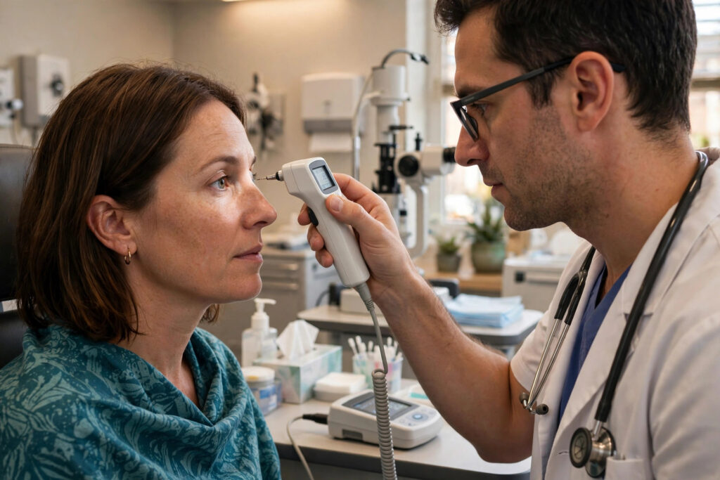
Corneal thickness is the gatekeeper measurement of LASIK candidacy. Before any laser touches your eye, your surgeon measures your cornea down to the micron and calculates whether your prescription can be corrected while leaving enough tissue behind for lifelong stability. Understanding this measurement helps you make sense of your screening results and your surgeon’s recommendations.
This article explains how corneal thickness is measured, what minimums surgeons look for, how your prescription strength changes the math, and what your options are if your measurements fall short. As with all refractive surgery topics, this is educational information, not medical advice; only a qualified refractive eye surgeon evaluating your own eyes can determine your candidacy.
Table of Contents
- How Corneal Thickness Is Measured
- Minimum Thickness Guidelines for LASIK
- The Residual Stromal Bed Rule
- How Prescription Strength Affects Tissue Removal
- What Happens If You Fall Short
- Protecting Long-Term Corneal Health
How Corneal Thickness Is Measured
The measurement is called pachymetry, and it is quick and painless. The most common method uses ultrasound: after numbing drops, a small probe touches the cornea for a second and reports the thickness in microns. Many modern clinics instead use non-contact optical methods, such as Scheimpflug imaging or optical coherence tomography, which map thickness across the entire cornea without touching the eye.
Full corneal mapping matters because the cornea is not uniformly thick; it is thinnest at the center and thicker toward the edges, and the thinnest point is not always dead center. A single central reading can miss an off-center thin spot, so reputable screenings produce a color-coded thickness map of the whole cornea. This map, combined with topography showing the cornea’s shape, gives the surgeon the complete picture.
Measurements are typically taken after you have been out of contact lenses for the required period, because lenses can temporarily alter corneal shape and hydration. Soft lens wearers are usually asked to remove them for several days to a couple of weeks before screening, and rigid lens wearers for longer. Accurate Eye Exam Cost Without Insurance in 2026“>eye exam measurements are the foundation of a safe surgical plan, so following the lens holiday matters.
Minimum Thickness Guidelines for LASIK
There is no single official minimum thickness for LASIK set by regulators; instead, surgeons work from the safety calculation described below. In practice, many surgeons like to see at least 500 microns of starting thickness for a standard LASIK candidate, and they grow cautious below that level. Average corneas measure roughly 540 to 560 microns at the center, so a large share of patients clear the bar comfortably.
But the starting number is only half the equation. A 505-micron cornea paired with a mild prescription may be a perfectly safe LASIK candidate, while a 560-micron cornea paired with a very high prescription might not be. This is why quoting a universal minimum is misleading, and why surgeons speak in terms of the residual bed, the tissue that remains after the procedure, rather than the starting thickness alone.
The FDA notes that patients with thin corneas may not be good LASIK candidates, and directs patients to discuss their individual measurements with their surgeon. If your screening report shows a number that worries you, ask the surgeon to walk through the full calculation rather than judging the raw figure on its own.
The Residual Stromal Bed Rule
The key safety concept is the residual stromal bed: the layer of untouched corneal tissue that must remain beneath the LASIK flap after the laser reshaping. A widely used guideline is to leave at least 250 to 300 microns of residual bed, with many surgeons preferring the higher end of that range or more. This untouched layer is what keeps the cornea structurally stable for decades.
The arithmetic is simple in concept: starting thickness, minus flap thickness, minus the tissue the laser will remove for your prescription, must leave at least the minimum residual bed. Modern femtosecond lasers create thinner, more predictable flaps than older mechanical blades, often around 100 microns, which has effectively lowered the thickness bar for LASIK over the years by consuming less tissue in the flap step.
Some surgeons also calculate the percent tissue altered, which expresses the combined flap and ablation depth as a percentage of total thickness. Lower percentages correlate with lower risk of post-operative weakening. Together, these calculations turn pachymetry from a single number into a personalized safety verdict. When the math works with margin to spare, you can proceed with confidence; when it does not, the alternatives are worth exploring.
How Prescription Strength Affects Tissue Removal
Every diopter of nearsightedness corrected requires removing a predictable depth of tissue, roughly a dozen or more microns per diopter depending on the treatment zone size. A mild prescription of minus two diopters might need only a shallow reshaping, while a minus eight prescription needs several times more. Astigmatism correction adds its own tissue demand on top.
The treatment zone, the diameter of the reshaped area, also matters. Larger optical zones give better night vision quality but remove more tissue, so surgeons balance the two. This is one reason very high prescriptions are harder to treat safely with LASIK: the tissue demand grows faster than most corneas can supply.
Farsightedness corrections remove tissue in a ring pattern rather than centrally, with their own calculation quirks. Whatever your prescription type, the surgeon should be able to tell you exactly how many microns your treatment requires. Patients who wear How to Put In Contact Lenses: A Beginner’s Guide“>contact lenses should know that long-term lens wear does not change these calculations, though the pre-screening lens holiday keeps measurements honest.
What Happens If You Fall Short
Failing the thickness calculation for LASIK is common and not a verdict on your eyes. The first alternative most surgeons discuss is PRK, which skips the flap entirely and therefore preserves the tissue LASIK would spend on one. Many patients who are poor LASIK candidates on thickness grounds are excellent PRK candidates, with equivalent long-term vision.
SMILE is a second possibility for borderline cases, since its small-incision approach preserves more of the cornea’s strong anterior layers than a LASIK flap. And when no corneal procedure fits safely, implantable collamer lenses bypass the cornea almost entirely, making them the go-to for thin corneas paired with high prescriptions.
What should not happen is a clinic agreeing to LASIK anyway by shaving the safety margins. The residual bed guideline exists because of real cases of post-LASIK ectasia, a progressive weakening and bulging of the cornea that can severely distort vision. It is rare in properly screened patients, which is precisely why screening standards must be respected. A surgeon who declines LASIK and offers alternatives is protecting you.
Protecting Long-Term Corneal Health
Whether or not you have laser surgery, corneal health deserves ongoing attention. After any refractive procedure, keep every scheduled follow-up so the surgeon can confirm the cornea is healing and stable. Report persistent blur, distortion, or halos promptly rather than waiting for the next appointment; early detection of any issue always beats late.
Everyday habits matter too. Do not rub your eyes vigorously, a habit linked to corneal stress over time. Wear quality sunglasses with full UV protection outdoors, since ultraviolet exposure contributes to several corneal and eye surface problems. Manage dry eye proactively with the drops or treatments your eye doctor recommends, because a healthy tear film supports the entire corneal surface.
Finally, keep copies of your screening measurements. If you ever seek a second opinion or consider an enhancement years later, having your original pachymetry and topography maps saves time and gives the next surgeon a baseline. Advances in UV Coating on Lenses Explained“>lens technology continue to improve non-surgical options as well, so whatever path your measurements point to, you have good choices. The National Eye Institute offers clear background reading on refractive errors and their correction.