
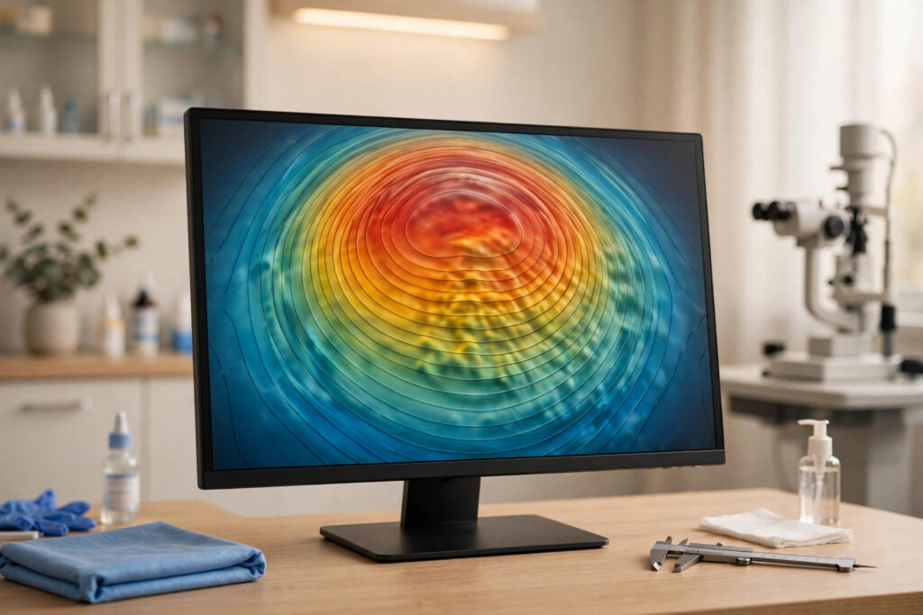
Topography-guided LASIK is a form of laser vision correction that customizes treatment using a highly detailed map of your cornea’s surface shape. Where conventional LASIK works from your glasses prescription alone, topography-guided treatment measures tens of thousands of points across the cornea and builds the laser plan from that anatomical data. The goal is a treatment shaped to your actual corneal terrain, including microscopic irregularities a standard prescription cannot capture.
This approach has gained significant traction in recent years and, by 2026, is offered alongside wavefront-guided treatments at many established refractive practices. This article explains what corneal topography measures, how a topography-guided procedure differs from wavefront-guided LASIK, who tends to be a good candidate, and what recovery looks like. Discuss your individual suitability with a qualified refractive eye surgeon before considering any procedure.
Table of Contents
- What Topography-Guided LASIK Is
- How Corneal Topography Mapping Works
- Topography-Guided vs Wavefront-Guided LASIK
- Ideal Candidates
- Treatment Day and Recovery
- Results, Limitations, and Cost
What Topography-Guided LASIK Is
The cornea provides roughly two-thirds of the eye’s focusing power, and its precise shape determines how cleanly light is focused. No cornea is a perfect dome — each has subtle elevations, depressions, and asymmetries unique to the individual. Corneal topography captures this landscape in fine detail, and topography-guided LASIK uses that map as the direct basis for the laser treatment.
In practical terms, the surgeon first acquires several high-quality topography scans of your cornea. Software analyzes the elevation data, compares it against an ideal corneal shape for your prescription, and calculates exactly where tissue must be removed to regularize the surface. The excimer laser then executes that plan with eye-tracking alignment, much like conventional LASIK — but guided by your corneal anatomy rather than your prescription alone.
The treatment has a regulatory history worth knowing. Topography-guided platforms received FDA approval for the treatment of myopia and astigmatism after clinical trials showed strong visual outcomes, and their approved indications have been the subject of ongoing study. In 2026, the technology is considered a mainstream premium option rather than an experimental one, available on current-generation laser platforms at many refractive centers.
It is useful to distinguish topography-guided treatment from topography as a diagnostic tool. Corneal topography is used in nearly every LASIK screening to rule out irregularities like keratoconus. Topography-guided LASIK goes a step further: it feeds the topographic data directly into the treatment itself. Every LASIK workup uses topography; only guided treatments let the map drive the laser.
How Corneal Topography Mapping Works
Modern corneal topographers, often based on Placido-disc or Scheimpflug imaging, capture the cornea’s shape in seconds. In a Placido-based system, you look at a target while illuminated concentric rings reflect off your tear film; a camera records the reflection pattern, and software reconstructs the surface from how the rings distort. Scheimpflug systems use rotating cameras to build a three-dimensional model that includes the cornea’s back surface and thickness as well.
The resulting maps are famously colorful — reds for steeper areas, blues for flatter ones — and experienced surgeons read them the way meteorologists read weather charts. For a guided treatment, though, the raw elevation data matters more than the colors: the software needs precise height measurements at thousands of points to compute the ablation profile.
Scan quality is everything. The tear film must be stable, because topography measures the tear film’s surface as a proxy for the cornea beneath it. Dry or unstable tears produce noisy maps, which is why clinics treat dry eye before scanning and may capture multiple maps to confirm consistency. Contact lens wear must be discontinued for the specified period beforehand, since lenses mold the cornea and the map must reflect its natural shape. A comprehensive How to Choose an Eye Doctor“>eye exam with careful ocular surface preparation underpins the whole process.
Once a clean map is acquired, planning software simulates the treatment and shows the surgeon the predicted post-operative corneal shape. The surgeon reviews and approves the plan — or adjusts it — before anything is transferred to the laser. This review step is where surgical judgment enters: the software proposes, but the surgeon disposes.
Topography-Guided vs Wavefront-Guided LASIK
Patients often ask which custom treatment is “better,” and the honest answer is that they measure different things. Wavefront aberrometry measures the eye’s total optical performance — how light travels through the cornea, lens, and everything in between — including higher-order aberrations from the whole system. Topography measures the cornea’s shape specifically, in exquisite anatomical detail.
Because the cornea dominates the eye’s optics, the two approaches often converge on similar treatments for typical eyes. They can diverge, however, when significant aberrations come from the crystalline lens rather than the cornea, or when the cornea has localized irregularities that wavefront measurements average over. Some surgeons view topography-guided treatment as especially logical for corneas with mild irregularities, since it regularizes the actual surface.
One practical difference lies in the treatment philosophy. Wavefront-guided LASIK aims to optimize the total optical system, including lens-derived aberrations. Topography-guided LASIK aims to create the most regular corneal surface possible for the target prescription, trusting that a clean corneal shape produces clean optics. Both philosophies have clinical support, and many surgeons choose based on the individual eye’s data rather than brand loyalty.
There is also a hybrid reality in modern practice: thorough clinics acquire both wavefront and topography data during screening, and the surgeon uses both to decide which guided treatment — or whether a non-LASIK procedure — best fits the eye. The maps inform each other. If you are comparing clinics, ask which measurements they take and how the surgeon uses them in planning, rather than focusing on a single technology name.
Ideal Candidates
The candidacy fundamentals match standard LASIK: adults with stable prescriptions, corneas of adequate thickness and healthy shape, no keratoconus or significant corneal disease, and realistic expectations. Within that group, topography-guided treatment is often discussed for patients whose corneal maps show mild irregularities or asymmetries that a conventional treatment would simply work around rather than address.
Patients who are particular about visual quality — night drivers, people bothered by glare or halos, those with visually demanding occupations — frequently ask about guided treatments, and topography-guided LASIK is a reasonable option to discuss. It is also sometimes considered for patients seeking enhancements after previous corneal surgery, where the corneal terrain is no longer pristine; the map-driven approach can regularize an already-treated surface in ways a prescription-based retreatment cannot.
There are boundaries. Topography-guided platforms carry specific approved treatment ranges for nearsightedness and astigmatism, and prescriptions outside those ranges need other solutions. Irregular corneas that suggest early keratoconus remain a contraindication for LASIK in any form. And as with all laser vision correction, the eyes must be free of uncontrolled dry eye, active infection, and healing disorders. If LASIK is ruled out, options like Bifocal vs Trifocal Lenses Explained“>eyeglasses with advanced optics remain excellent non-surgical alternatives.
The screening appointment is the real decision point. Expect repeated topography scans, wavefront measurements, corneal thickness mapping, pupil sizing, and a dilated retinal exam. A clinic that rushes this workup — or one that recommends a guided premium treatment without showing you your own maps — deserves a second opinion.
Treatment Day and Recovery
The procedure itself mirrors conventional LASIK: numbing drops, flap creation, the excimer laser applying the topography-derived treatment, and flap repositioning. The treatment may take slightly longer than a conventional ablation because the customized profile can involve more laser pulses, but the difference is measured in seconds, and the overall experience is the same.
Recovery follows the standard LASIK arc. Vision is hazy immediately afterward, improves markedly within hours, and is typically functional by the next morning’s check-up. Most patients return to desk work within a day or two. Prescribed drops — usually an antibiotic and a steroid taper — plus frequent lubrication support healing, and surgeons advise against eye rubbing, swimming, and eye makeup for the period they specify.
Over the following weeks, vision continues to refine as the cornea settles into its new shape and the tear film stabilizes. Because topography-guided treatment aims for a highly regular corneal surface, many patients report clean, crisp vision with good night-driving comfort once healing completes. Follow-up visits confirm the outcome and give the surgeon a chance to address any residual dryness or fluctuation.
Protecting the investment matters: quality When to Replace Your Sunglasses: Signs to Watch“>sunglasses with full UV protection are sensible after any corneal procedure, and ongoing routine eye care continues on its normal schedule. LASIK reshapes the cornea; it does not exempt you from future eye health monitoring.
Results, Limitations, and Cost
Published clinical data for topography-guided LASIK has shown strong outcomes, with high proportions of patients achieving excellent uncorrected vision and reporting good quality of vision. Surgeons who use the technology regularly often describe particular satisfaction among patients with pre-existing corneal irregularities, who may notice improvements in visual quality beyond what their prescription change alone would predict.
The limitations deserve equal airtime. The treatment is only as good as the topographic data, and poor tear film or inconsistent scans can compromise the plan — which is why meticulous clinics repeat scans and optimize the ocular surface first. The approved treatment range is narrower than some wavefront platforms, so higher prescriptions may not qualify. And topography-guided LASIK remains LASIK: it needs adequate corneal thickness, cannot fix presbyopia’s underlying mechanism, and carries the standard profile of surgical risks.
Cost typically sits at the premium end of the LASIK spectrum, reflecting the advanced diagnostics and planning involved. As with any refractive procedure, compare what the quoted price includes — screening, treatment, follow-up visits, and the enhancement policy — rather than the headline number alone. Prices far below the local norm, or pressure to decide on the spot, are warning signs regardless of the technology.
For neutral background reading, the FDA’s LASIK surgery expectations page and the National Eye Institute’s refractive errors overview are trustworthy starting points. Then bring your questions — and your corneal maps — to a qualified refractive eye surgeon for advice tailored to your eyes.