
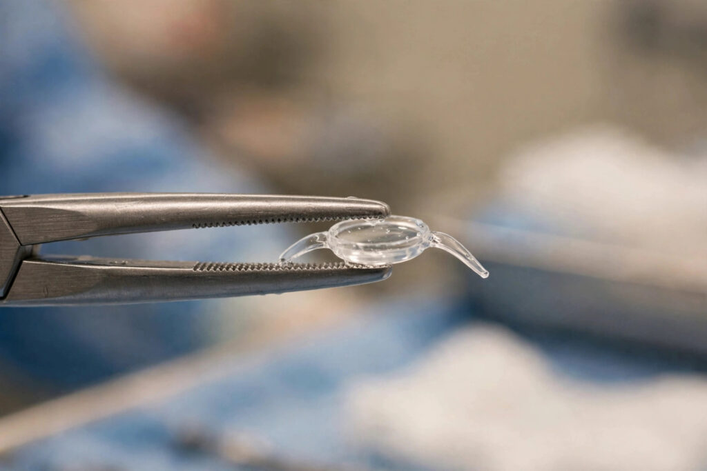
ICL surgery, formally known as implantation of a phakic intraocular lens, corrects vision in a fundamentally different way from LASIK, PRK, or SMILE. Instead of reshaping the cornea with a laser, the surgeon places a thin, flexible artificial lens, made from a biocompatible material called collamer, inside the eye in front of the natural lens. The cornea is left essentially untouched.
Because nothing is removed from the eye, ICL is reversible in a way laser procedures are not: the lens can be taken out if ever needed. It has become an important option for patients with very high prescriptions or thin corneas who are not candidates for laser vision correction. This guide explains how ICL eye surgery works, who it suits, what recovery involves, and what to weigh before deciding. Discuss every step with a qualified refractive eye surgeon experienced in lens-based procedures.
Table of Contents
- What the Implantable Collamer Lens Is
- How ICL Surgery Works
- ICL vs Laser Vision Correction
- Who Is a Good Candidate
- Recovery Timeline
- Risks and Long-Term Outlook
- Cost and Final Thoughts
What the Implantable Collamer Lens Is
The implantable collamer lens is a soft, flexible lens made from collamer, a material combining collagen with a polymer that the eye tolerates well. It is custom manufactured to the patient’s prescription and eye measurements, including versions that correct astigmatism. The lens sits in the posterior chamber of the eye, behind the iris and in front of the natural crystalline lens, where it stays permanently unless removed.
Modern ICL designs include a tiny central port that allows natural fluid circulation inside the eye, which removed the need for the preventive laser iridotomy that earlier generations required. The lens also provides built-in UV protection. Because it works inside the eye rather than on the cornea, it can correct a wider range of nearsightedness than laser procedures, including very high prescriptions.
Unlike a contact lens, the ICL cannot be felt once implanted and requires no daily maintenance. Unlike laser surgery, it adds tissue rather than removing it, which is the basis for both its reversibility and its suitability for eyes that laser cannot safely treat.
How ICL Surgery Works
ICL surgery is performed as an outpatient procedure under numbing drops, sometimes with light sedation. The surgeon makes a tiny incision at the edge of the cornea, typically only a few millimeters wide, and injects the folded collamer lens through it. Inside the eye, the lens unfolds and is gently positioned into place behind the iris.
The procedure usually takes around twenty to thirty minutes per eye, and many surgeons treat one eye at a time with a short interval between eyes. Patients are awake throughout and generally report feeling pressure rather than pain. The small incision is self-sealing and does not require stitches.
Because the cornea is not reshaped, vision often improves remarkably quickly, with many patients noticing sharp vision within hours. Antibiotic and anti-inflammatory drops are used for a few weeks afterward, and follow-up visits confirm the lens is sitting correctly and eye pressure is normal.
ICL vs Laser Vision Correction
The most important difference is reversibility. Laser procedures permanently remove corneal tissue, while an ICL can be removed or exchanged if vision needs change significantly or if any issue arises. For younger patients whose prescriptions might still evolve, that flexibility is a meaningful advantage worth discussing.
ICL also sidesteps the dry eye concern associated with laser surgery. Since the corneal nerves are not disrupted by a flap or surface ablation, post-operative dryness is generally less of an issue, which matters for patients who already struggle with dry eye while wearing Colored Contact Lenses: Safety Guide and Tips“>contact lenses.
On the other side, ICL is intraocular surgery, which carries a different risk profile from surface laser work, including small risks of elevated eye pressure, cataract formation over time, and infection inside the eye. It is also generally more expensive than laser procedures. Neither option is universally better; the right choice depends on prescription, corneal anatomy, age, and personal priorities.
Who Is a Good Candidate
ICL’s classic candidate is the adult with moderate to very high nearsightedness, with or without astigmatism, whose corneas are too thin for safe laser ablation or whose prescription exceeds what lasers can treat. Patients with significant dry eye who are poor laser candidates are also commonly evaluated for ICL.
Anatomy matters as much as prescription. The eye needs adequate depth in the anterior chamber to accommodate the lens safely, and the endothelial cell count, the cells keeping the cornea clear, must be healthy. Precise measurements with specialized imaging determine whether the lens will fit correctly.
General requirements mirror other refractive procedures: stable vision, healthy eyes without glaucoma or uncontrolled disease, and no active eye infection. A comprehensive Retinal Imaging (Fundus Photography) Explained“>eye exam with anterior segment imaging is the gateway step, and anyone considering ICL should seek a surgeon who performs lens-based procedures regularly.
Recovery Timeline
Recovery after ICL surgery is typically quick and comfortable. Many patients see clearly within hours of the procedure, and most return to desk work within a day or two. The eye may feel mildly scratchy or watery on the first day, which usually settles fast.
Prescribed drops are used for several weeks to prevent infection and control inflammation, and follow-up visits in the first days and weeks check lens position and eye pressure. Strenuous exercise, swimming, and eye rubbing are restricted for the period the surgeon specifies, similar to other eye surgeries.
Because there is no corneal healing wound to speak of, visual fluctuations are usually minor after the first days. Long-term, the lens simply stays in place doing its job, with routine annual eye examinations to monitor eye health as with any eyes.
Risks and Long-Term Outlook
ICL surgery has a strong safety record in properly selected patients, but honest counseling includes the risks. Elevated intraocular pressure can occur in the early period and is monitored at follow-ups. There is a small long-term risk of cataract formation since the lens sits close to the natural lens, and rare risks of infection or lens malposition that might require repositioning.
Endothelial cell loss, the gradual decline of the cornea’s inner cell layer, is tracked over years, which is why adequate pre-operative cell counts and ongoing monitoring matter. Night vision symptoms such as halos can occur but are generally reported less often than after some laser procedures.
The long-term outlook is reassuring: the collamer material is designed to remain biocompatible indefinitely, and the lens does not degrade or need scheduled replacement. If the patient’s vision changes substantially decades later, the lens can be exchanged or removed and cataract surgery performed normally when age makes it necessary.
Cost and Final Thoughts
ICL surgery typically costs more than LASIK or PRK, reflecting the custom-manufactured lens, the intraocular surgical setting, and the specialized expertise involved. Pricing varies by region and practice, so compare what is included: measurements, the lens itself, surgery, medications, and follow-up care.
ICL deserves serious consideration from anyone told they are not a laser candidate because of thin corneas or a very high prescription. It is also worth discussing for patients with significant dry eye or those who value reversibility. At the same time, excellent Progressive Lenses vs Bifocals: Pros and Cons“>eyeglasses with modern high-index lenses remain a perfectly good choice, and quality sunglasses with UV protection are wise for every post-operative eye.
Take time choosing both the procedure and the surgeon, ask about the surgeon’s ICL-specific volume and complication rates, and read the FDA’s information on phakic intraocular lenses as a neutral reference. The American Academy of Ophthalmology’s overview is another solid source before your consultation.